Our lab is dedicated to making advancements in biomedical diagnostics and imaging. Our research spans a variety of topics including, spectroscopic analysis of bio molecules on the cellular and molecular levels, deep tissue imaging, machine learning, and computer aided diagnostics. We have collaborative relationships with the Air Force Research Lab, University of Sao Paulo, and various research groups here at Texas A&M University.
Students interested in gaining research experience, and researchers interested in collaboration are encouraged to contact us.
People
The researchers in this lab are under the direction of Dr. Vladislav V. Yakovlev. We have numerous projects, present at conferences, and publish regularly in high quality journals. The People link will guide you to discover more information about us.
Research
Displayed here are some featured projects of the lab. For a more comprehensive list of our ongoing work please follow the Research link.
Retinal Laser Lesion Segmentationby Eddie Gil Healthcare providers face a data deluge. This makes quick and efficient decision making challenging. Having automated assist systems can alleviate this issue and save lives. Computer aided diagnostics describes techniques to assist medical professionals not replace them. One such example is in retinal lesion segmentation. High power lasers have become easily accessible. Retinal Laser damage may occur under a variety of conditions. However, identifying retinal lesions is challenging and typically requires a panel of ophthalmologists. We trained a fully convolutional network to perform automated segmentation of retinal laser lesions in fundus images. We trained a similar network to perform semantic segmentation of the images as well to display whether damage was, photothermal, photomechanical, or photo chemical. 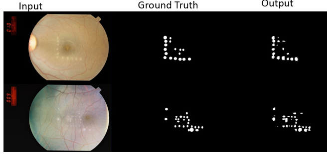 Fundus images input to the segmentation network alongside their groudtruths and the network output.  Preprocessed image alongside semantic segmentaiton result from network. Color in the segmentation image maps to photothermal, photomechanical, and photochemical damage. |
Traumatic Brain InjuryService members experience traumatic brain injury (TBI) at a rate between 24-41% per 10,000 soldiers-years. We aim to address knowledge gaps in order to develop better treatments for TBI. The knowledge gap in our current understanding of TBI comes from biomechanical changes at the tissue and cellular levels. Brillouin spectroscopy can be performed in vivo to extract mechanical information at these scales without need to euthanize the animal test subject afterwards. This makes Brillouin spectroscopy an exciting and ideal modality for addressing this knowledge gap. 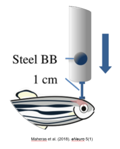 A diagram showing a scheme for inducing traumatic brain injury in zebrafish using a steel ball bearing. This set up is discussed in Maheras etal. (2018) eNeuro 5(1) 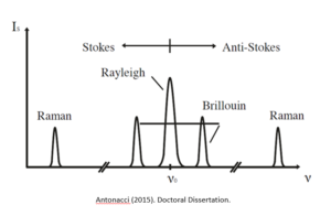 A diagram of Brillouin spectroscopy peaks from Antonacci (2015). Doctoral Dissertation
|
Brillouin Scatteringby Sean O’Connor and Dominik Doktor Understanding physiological mechanisms at the organelle level is crucial to the development of novel treatments. Viscoelastic properties of materials are of particular interest. Impulsive Stimulated Brillouin Scattering (ISBS) is an emerging nonlinear spectroscopy technique which can be used to probe these viscoelastic properties. The advantage of this technique is that it requires less acquisition time than Spontaneous Brillouin Scattering ( SpBS). Our focus is to improve the spatial resolution of ISBS systems to cellular and organelle level imaging. This can later be used to reveal insights about mitochondria, cytoskeletons, etc.
 Schematic for laser system 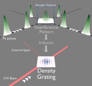 Schematic for optical phenomena. Ballmann (2017). Doctoral Dissertation |
Compressed Hyperspectral Ramanby Mark Keppler Hyperspectral microscopy captures spatial information about a scene in a series of images, where each image covers a different wavelength over a large portion of the electromagnetic spectrum. Key Points:
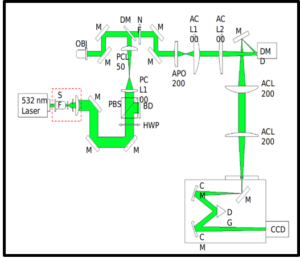 Schematic of the hyperspectral raman imaging system. 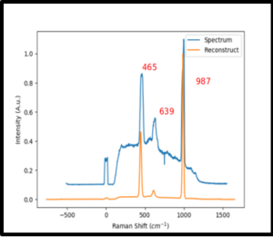 Barium Sulfate Spectrum acquired from 1 line in the hyperspectral raman image. 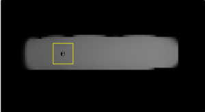 resulting 2D reconstruction of acquired image. |
Publications
Shown are some publications of interest.
Assessment of Local Heterogeneity in Mechanical Properties of Nanostructured Hydrogel Networks
Enhanced coupling of light into a turbid medium through microscopic interface engineering
Towards optical brain imaging: getting light through a bone
Enhanced second harmonic generation efficiency via wavefront shaping
News
Here are some news stories about our lab!

