Retinal Laser Lesion Segmentationby Eddie Gil Healthcare providers face a data deluge. This makes quick and efficient decision making challenging. Having automated assist systems can alleviate this issue and save lives. Computer aided diagnostics describes techniques to assist medical professionals not replace them. One such example is in retinal lesion segmentation. High power lasers have become easily accessible. Retinal Laser damage may occur under a variety of conditions. However, identifying retinal lesions is challenging and typically requires a panel of ophthalmologists. We trained a fully convolutional network to perform automated segmentation of retinal laser lesions in fundus images. We trained a similar network to perform semantic segmentation of the images as well to display whether damage was, photothermal, photomechanical, or photo chemical. 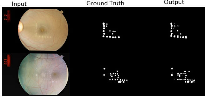 Fundus images input to the segmentation network alongside their groudtruths and the network output.  Preprocessed image alongside semantic segmentaiton result from network. Color in the segmentation image maps to photothermal, photomechanical, and photochemical damage. |
Traumatic Brain InjuryService members experience traumatic brain injury (TBI) at a rate between 24-41% per 10,000 soldiers-years. We aim to address knowledge gaps in order to develop better treatments for TBI. The knowledge gap in our current understanding of TBI comes from biomechanical changes at the tissue and cellular levels. Brillouin spectroscopy can be performed in vivo to extract mechanical information at these scales without need to euthanize the animal test subject afterwards. This makes Brillouin spectroscopy an exciting and ideal modality for addressing this knowledge gap. 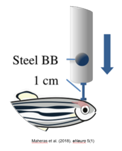 A diagram showing a scheme for inducing traumatic brain injury in zebrafish using a steel ball bearing. This set up is discussed in Maheras etal. (2018) eNeuro 5(1) 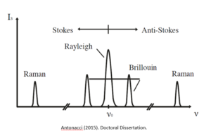 A diagram of Brillouin spectroscopy peaks from Antonacci (2015). Doctoral Dissertation
|
Brillouin Scatteringby Sean O’Connor and Dominik Doktor Understanding physiological mechanisms at the organelle level is crucial to the development of novel treatments. Viscoelastic properties of materials are of particular interest. Impulsive Stimulated Brillouin Scattering (ISBS) is an emerging nonlinear spectroscopy technique which can be used to probe these viscoelastic properties. The advantage of this technique is that it requires less acquisition time than Spontaneous Brillouin Scattering ( SpBS). Our focus is to improve the spatial resolution of ISBS systems to cellular and organelle level imaging. This can later be used to reveal insights about mitochondria, cytoskeletons, etc.
 Schematic for laser system 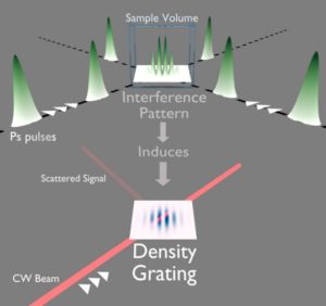 Schematic for optical phenomena. Ballmann (2017). Doctoral Dissertation. Visualization of transient grating produced from intersecting pump pulses followed by a probe which diffracts off of the induced grating. |
Compressed Hyperspectral Ramanby Mark Keppler Hyperspectral microscopy captures spatial information about a scene in a series of images, where each image covers a different wavelength over a large portion of the electromagnetic spectrum. Key Points:
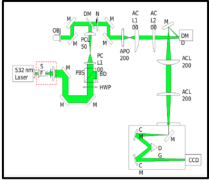 Schematic of the hyperspectral raman imaging system. 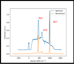 Barium Sulfate Spectrum acquired from 1 line in the hyperspectral raman image. 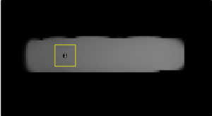 resulting 2D reconstruction of acquired image. |
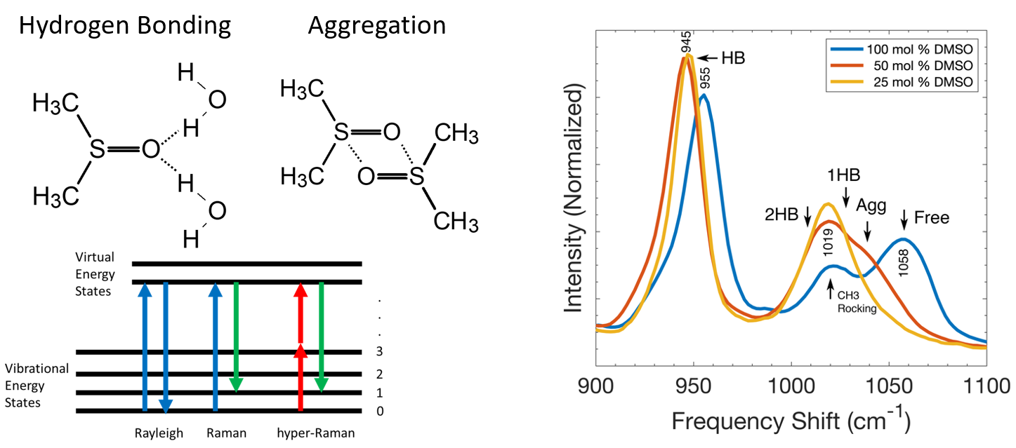
Hyper Raman Spectroscopy to Study Biomolecule interactionsChristopher Marble and Xingqi Xu The vibrational modes of biologically important molecules including proteins, nucleic acids and lipids provide insight into their structure, as well as means to study the metabolic processes and biomarker expression of cells. Raman spectroscopy is an essential optical tool for detecting the vibrational modes of biomolecules. Using a custom built 8 ps laser system, we are exploring the application of hyper-Raman scattering as a complementary technique that allows for detection of vibrational modes that are silent under Raman excitation. Beyond studying the hyper-Raman spectra of biomolecules, we are also using hyper-Raman scattering to probe fundamental questions of biology such as how hydrogen bonding between water and polar functional groups of biomolecules modifies the existing “network” of water-water hydrogen bonding as well as how water solvates biomolecules by breaking up intermolecular bonds between biomolecules in water poor areas of the cell.
|
|

