• Skip to primary navigation
  • Skip to main content
  • Home
  • Research
  • People
  • Courses
  • News
  • Publications
  • Resources
  • Facility

Advanced Spectroscopy Laboratory

Texas A&M University College of Engineering

Uncategorized

Yakovlev Looking To Shed Light Deeper Into The Human Brain

Posted on September 18, 2017 by John Paul Hernandez Alcala

Original Story Date: July 28, 2017
By Marcus Misztal, Texas A&M University College of Engineering

The inner workings of the human brain have always been a subject of great interest. Unfortunately, it is fairly difficult to view brain structures or intricate tissues due to the fact that the skull is not transparent by design. The reality is that light scattering is the major obstacle for deep penetration into tissue.

Dr. Vladislav Yakovlev, professor in the Department of Biomedical Engineering at Texas A&M University, has been developing a more efficient way of propagating light through an opaque medium. Propagation of light refers to the way that light travels from one point to another, in this case, through a medium, such as human tissue.

The new method involves making a minimally invasive hole within the medium, which is smaller in diameter than needles that are currently being used within the medical field. The process shows a great deal of promise in many uses, including viewing brain structure through the skull and imaging blood through skin tissue.

This technology has the potential to be used to better understand in living patients diseases like Chronic Traumatic Encephalopathy (CTE), Alzheimer’s and other brain diseases that currently can only be studied post-mortem.

The technology could even be extended outside the realm of biomedical engineering to develop a more efficient way of seeing through fog while driving. This can be accomplished by deploying a laser pulse that could be sent through fog and evaporate water. This would allow drivers to have a safer experience during hazardous driving conditions and would work exactly as the method used in biomedical engineering applications.

Vladislav Yakovlev has been developing a more efficient way of propagating light through an opaque medium.

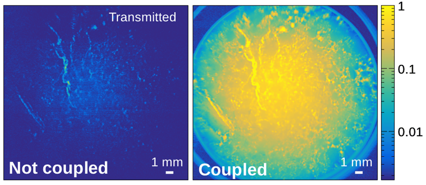
The holes used to pass the light through are a few hundred micrometers in depth and a width of 20 to 30 microns. A micron is one millionth of a meter, and by comparison a single strand of human hair is about 75 microns in diameter. The light is then coupled into the opaque material resulting in an increase of magnitude of optical transmission into the material. The material that light is passed through is also referred to as the scattering medium.

The report documenting the work of Yakovlev was recently published in Proceedings of the National Academy of Sciences of the United States of America and definitively demonstrated that light injected into the scattering medium will remain there for an extended period of time. The amount of time that the photons remained was increased by a factor of 100.

One of the challenges facing researchers is that of optical absorption within tissues. However, because the new method is wavelength independent, the wavelength can be specified to perform measurements in a specific part of the light spectrum. This approach has the potential to yield analytical information about the composition and structure of the medium or tissue.

The project was a collaborative effort between Dr. Brian Applegate and Dr. Javier Jo, professors in the biomedical engineering department at Texas A&M. The observed data from the project was a collaborative process with Yale University and Missouri University of Science.

The research was sponsored by the Department of Defense and the National Science Foundation.

###

This story by Marcus Misztal originally appeared on the College of Engineering website.


Filed Under: Uncategorized

New Technology Developed at Texas A&M Could Improve Diabetes Management

Posted on June 30, 2017 by John Paul Hernandez Alcala

A newly developed method for detecting glucose based on how it absorbs a specific type of light could spell the end of the painful, invasive finger-prick tests diabetics rely on to monitor their condition, says a Texas A&M University biomedical engineer who is developing the technology.

Using optical technology that essentially sends a twisting, directional type of light at a glucose-containing sample, a team of researchers led by Vladislav Yakovlev, professor in Texas A&M’s Department of Biomedical Engineering, has been able to accurately detect glucose concentrations by measuring how glucose absorbs this light at a molecular level. His findings may translate into a more effective means of diabetes management for the millions suffering from the disease.

The research, which was spearheaded by undergraduate student Carlos Tovar, under the guidance of Yakovlev and graduate students Brett Hokr and Zhaokai Meng, was presented at this year’s SPIE Photonics West conference. Authorities on biophotonics, nanophotonics and biomedical optics from throughout the world convene annually at the high-tech conference to discuss their cutting-edge work. Each year, the conference attracts more than 20,000 people who want to see, learn about and purchase the latest devices, components and systems that are driving trends such as state-of-the art medical technologies, smart manufacturing and autonomous vehicles.

Yakovlev-glucose detectionYakovlev, an authority on biomedical diagnostics and imaging instrumentation, says standard glucose-monitoring techniques such as finger-prick methods are somewhat of a guessing game because they cannot achieve continuous monitoring of a patient’s blood-glucose levels. What’s more, patients are advised to self-administer these tests at least three times a day, but because of the painful and awkward nature of the tests many people don’t comply with this instruction, he notes.

The result is inadequate and, at times, ineffective management of the disease, he says, and it’s a growing problem given that diabetes is reaching epidemic levels. In the United States alone, the disease affects 29.1 million people and is the seventh-leading cause of death. Sobering statistics such as these, Yakovlev notes, demonstrate a clear need for a noninvasive, pain-free and accurate technique to monitor blood-glucose levels.

That need could be answered by the optical detection technology being developed by Yakovlev and his team at Texas A&M. Though it’s still in its infancy, this technology might one day be implemented in devices such as smart watches and bracelets where it would provide patients with hassle-free, continuous monitoring and alerts when their blood-glucose levels slip to dangerous levels. For now, the technology is being refined in the laboratory with the hopes of one day moving to human trials if results continue to be positive, Yakovlev says.

The technology basically works by measuring how individual glucose molecules absorb a specific wavelength of light that is characterized as right and left circular polarized, Yakovlev explains. In very simple terms, this is light that twists to the right and light that twists to the left, he explains. By measuring the specific manner in which glucose molecules vibrate and absorb this light, Yakovlev is able to differentiate the molecules from their surroundings and calculate glucose concentrations.

It’s a small but measurable effect, he says, that is due to a geometric property of the glucose molecule known as chirality. Glucose’s chiral structure, which is a result of its arrangement of atoms, means that its molecule cannot be superimposed over its mirror image. For non-engineers that concept may be difficult to imagine, but think of a pair of shoes. The left shoe is a mirror image of the right shoe, but they are not interchangeable. That same property exists for certain molecules such as glucose, and it results in the molecule absorbing light in a specific way, Yakovlev explains.

This specificity, he notes, enables him to discriminate glucose molecules from other surrounding biological molecules – something that historically has proven difficult with other detection approaches. For example, Yakovlev explains, the large presence of water in tissue has typically created a problem when it comes to attempting to detect glucose with optical technology because water also absorbs light and in doing so masks the presence of glucose. In addition, protein molecules can appear similar, necessitating the factoring in of additional variables into detection technology in order to obtain reliable data.

These issues are known as calibration problems, he says. However, Yakovlev’s technology overcomes these obstacles by reaching the proper penetration depths and producing a unique, measurable effect that, in a sense, removes the background noise from the picture, leaving behind the glucose signal.


“This technology has the potential to separate the presence of glucose from its surroundings, avoiding the calibration problem while circumventing the huge absorption of water in the fundamental vibrational region of the spectrum, allowing for clinically relevant penetration depths in biological tissue,” Yakovlev says.


SPIE, the international society for optics and photonics, was founded in 1955 to advance light-based technologies. Serving more than 264,000 constituents from approximately 166 countries, the not-for-profit society advances emerging technologies through interdisciplinary information exchange, continuing education, publications, patent precedent and career and professional growth.

About the Department of Biomedical Engineering
Committed to solving the world’s greatest health problems through the exploration of new ideas, integrated research and innovation, the Department of Biomedical Engineering at Texas A&M is producing the next generation of biomedical engineers, developing new technologies and new jobs, and achieving revolutionary advancements for the future of health care. The department has unique strengths in regenerative engineering, medical augmentation, molecular diagnostics/theranostics, tele-health, and precision medicine, and its faculty members are internationally recognized with collaborative relationships that span engineering, physical and natural sciences, medicine and veterinary sciences.

-30-

Contact: Professor Vladislav Yakovlev at (979) 458-2326 or via email at yakovlev@tamu.edu or Ryan A. Garcia at (979) 979.847.5833 or via email at ryan.garcia99@tamu.edu.

Original story can be found here: http://engineering.tamu.edu/news/2016/06/22/glucose-detection

Filed Under: Uncategorized

New Technology Could Mean Better Chemical Analysis on Earth and in Space

Posted on June 30, 2017 by John Paul Hernandez Alcala

A new lightweight, energy-efficient tool for analyzing a material’s chemical makeup could improve the detection abilities of various technologies, ranging from bomb-detecting drones to space rovers searching for signs of life, says a Texas A&M University biomedical engineer who is part of the team developing the instrument.

The tool makes use of optical communications fiber to collect and transmit light as it interacts with the material being studied, explains Vladislav Yakovlev, professor in the Department of Biomedical Engineering at Texas A&M. Compared with conventional technology, the newly designed measurement system is 95 percent lighter, requires 65 percent less energy and is only about a third of the cost, he says. The system is detailed in the latest issue of the scientific journal Proceedings of the National Academy of Sciences.

Perhaps just as important, because of the way in which the system is constructed, it’s significantly sturdier than current technology, Yakovlev notes. This increased robustness, coupled with a massive reduction in the system’s overall weight and decreased energy requirements, makes the technology a prime candidate for integration into lightweight unmanned aircraft vehicles used for remotely sensing explosives, he says. But Yakovlev’s technology is not limited to terrestrial applications; those same attributes make the system an ideal tool for use on space-based vehicles where mechanical shocks and excessive vibrations associated with the launchings and landings have often damaged analysis technologies, Yakovlev notes.

Yakovlev is improving Raman spectroscopy[By using a simple optical fiber, Professor Vladislav Yakovlev is eliminating the need for bulky and costly spectrometers (shown on his left).]

In essence, Yakovlev’s system is a “better mousetrap” – a reinvention of a technology known as Raman spectroscopy. Raman spectroscopy is a widely used, nondestructive method for performing chemical analysis of a material. It involves analyzing a spectrum of light as it interacts with the molecules of a material. During this process, researchers shine light – typically a laser – on a material. As that light interacts with the molecules of that material, it scatters and changes color in a unique way, depending on the material itself. Because the resulting spectrum is unique to the material, it serves as a sort of “fingerprint” by which researchers can identify the exact chemical composition of that material.

However, the Raman effect is too weak to see with the naked eye, so researchers use a device known as a spectrometer to collect and analyze the scattered light. Although extremely effective, spectrometers, Yakovlev explains, are costly, delicate and require large amounts of energy to power them – all characteristics that limit their use in extreme conditions.

With this in mind, Yakovlev and his team went about building a lightweight and energy-efficient Raman measurement system that did not require a spectrometer.  The fiber-based setup they developed, Yakovlev explains, is capable of highly sensitive signal detection by using optical fibers to collect and transmit light as opposed to allowing the light to pass through lenses, as is the case in conventional Raman spectroscopy. The network of fibers, he adds, can be securely and rigidly attached to a small detector (a device known as a photomultiplier), enabling the entire apparatus to become resistant to mechanical shocks because there are no moving parts.

“We looked at how you disperse light and how you then analyze light,” Yakovlev said. “Instead of using the traditional spectrometer we are using an optical communications fiber to collect and transmit the light. Then we use a standard detector that is commonly used in biomedical and military applications to analyze the arrival time of photons. The detector has a short time response, so it’s fast, and there is no need for a spectrometer. Any application that involves using such a device for a long period of time with no access to an energy source would benefit from our technology.”

Joining Yakovlev in this research effort are Professor Marlan O. Scully of Texas A&M, Princeton University and Baylor University; Associate Professor Javier Jo of Texas A&M; Professor Kevin K. Lehmann of the University of Virginia; Texas A&M Engineering Experiment Station Research Engineer Georgi I. Petrov and Shuna Cheng and graduate student Zhaokai Meng of Texas A&M.

About the Department of Biomedical Engineering

Committed to solving the world’s greatest health problems through the exploration of new ideas, integrated research and innovation, the Department of Biomedical Engineering at Texas A&M is producing the next generation of biomedical engineers, developing new technologies and new jobs, and achieving revolutionary advancements for the future of health care. The department has unique strengths in regenerative engineering, medical augmentation, molecular diagnostics/theranostics, tele-health, and precision medicine, and its faculty members are internationally recognized with collaborative relationships that span engineering, physical and natural sciences, medicine and veterinary sciences.

-30-

Contact: Professor Vladislav Yakovlev at (979) 458-2326 or via email at yakovlev@tamu.edu or Ryan A. Garcia at (979) 979.847.5833 or via email at ryan.garcia99@tamu.edu.

Original link can be found here: http://engineering.tamu.edu/news/2015/09/23/better-raman

Filed Under: Uncategorized

New Material developed at Texas A&M Could Improve Ultrasound Technology

Posted on June 30, 2017 by John Paul Hernandez Alcala

Ultrasound technology could soon experience a significant upgrade that would enable it to produce high-quality, high-resolution images thanks to the development of a new key material by a team of researchers that includes a professor in Texas A&M University’s Department of Biomedical Engineering.

The material, which converts ultrasound waves into optical signals that can be used to produce an image, is the result of a collaborative effort by Texas A&M Professor Vladislav Yakovlev and researchers from King’s College London, The Queen’s University of Belfast and the University of Massachusetts Lowell. Their findings appear in the current issue of Advanced Materials.

Photo of Dr. Vladislav Yakovlev

(Pictured above: Professor Yakovlev examines a prototype of the transducer, which converts ultrasound waves into optical signals.)

The engineered material, known as a “metamaterial,” offers significant advantages over conventional ultrasound technology, which generates images by converting ultrasound waves into electrical signals, Yakovlev explains. Although that technology has advanced throughout the years — think of the improvement in sonogram images — it is still largely constrained by bandwidth and sensitivity limitations, he says. These limitations, he adds, have been the chief obstacle when it comes to producing high-quality images that can serve as powerful diagnostic tools.

The metamaterial developed by Yakovlev and his colleagues is not subject to those limitations, primarily because it converts ultrasound waves into optical signals rather than electrical ones. The optical processing of the signal does not limit the bandwidth or sensitivity of the transducer (converter) — and that’s important for producing highly detailed images, Yakovlev says.

“A high bandwidth allows you to sample the change of distance of the acoustic waves with a high precision,” Yakovlev notes. “This translates into an image that shows greater detail. Greater sensitivity enables you to see deeper in tissue, suggesting we have the potential to generate images that might have previously not been possible with conventional ultrasound technology.”

In other words, this new material may enable ultrasound devices to see what they haven’t yet been able to see. That advancement could significantly bolster a technology that is employed in a variety of biomedical applications. In addition to being used for visualizing fetuses during routine and emergency care, ultrasound is used for diagnostic purposes in incidents of trauma and even as a means of breaking up tissue and accelerating the effects of drugs therapies.

Illustration of optical signal

(Pictured right:  An optical signal, represented by the red arrow, comes into contact with the metamaterial and interprets the ultrasound waves, resulting  in an altered optical signal that is processed to produce a high-quality image.)

While Yakovlev’s research is not yet ready for integration into ultrasound technology, it has successfully demonstrated how conventional technology can be substantially improved by using the newly engineering material created by his team, he notes.

The material, he notes, consists of golden nanorods embedded in a polymer known as polypyrolle. An optical signal is sent into this material where it interacts with and is altered by incoming ultrasound waves before passing through the material. A detection device would then read the altered optical signal, analyzing the changes in its optical properties to process a higher resolution image, Yakovlev explains.

“We developed a material that would enable optical signal processing of ultrasound,” Yakovlev says. “Nothing like this material exists in nature so we engineered a material that would provide the properties we needed. It has greater sensitivity and broader bandwidth. We can go from 0-150 MHz without sacrificing the sensitivity. Current technology typically experiences a substantial decline in sensitivity around 50 MHz.

“This metamaterial can efficiently convert an acoustic wave into an optical signal without limiting the bandwidth of the transducer, and its potential biomedical applications represent the first practical implementation of this metamaterial.”

Yakovlev’s collaborators are Wayne Dickson and Anatoly Zayats of King’s College London; John McPhillips, Antony Murphy and Robert Pollard of The Queen’s University of Belfast; and Viktor Podolskiy of the University of Massachusetts Lowell.


Contact: Professor Vladislav Yakovlev at (979) 458-2326 or via email at yakovlev@tamu.edu.

Original article link found here: http://engineering.tamu.edu/news/2013/03/06/new-material-developed-at-texas-am-could-improve-ultrasound-technology

Filed Under: Uncategorized

© 2016–2025 Advanced Spectroscopy Laboratory Log in

Texas A&M Engineering Experiment Station Logo
  • State of Texas
  • Open Records
  • Risk, Fraud & Misconduct Hotline
  • Statewide Search
  • Site Links & Policies
  • Accommodations
  • Environmental Health, Safety & Security
  • Employment